PRODUCTS
NIRD®– HI (Hematoma Imager)
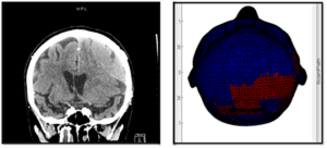
This device is designed to image the presence of intracranial bleeds within 1-3 minutes. The underlying technology is designed to specifically see blood – abnormal volumes of blood that should not be present in a healthy and normal head. Our novel approach incorporates motion-tracking and shape-recovery algorithms, and extremely high sample rate, allowing us to provide and image that very closely corresponds to a CT scan without the requirement of an expert to interpret the image. This device is meant to be used as an adjunct to CT scans (the Gold standard) and is not meant to be used in place of CT scans.
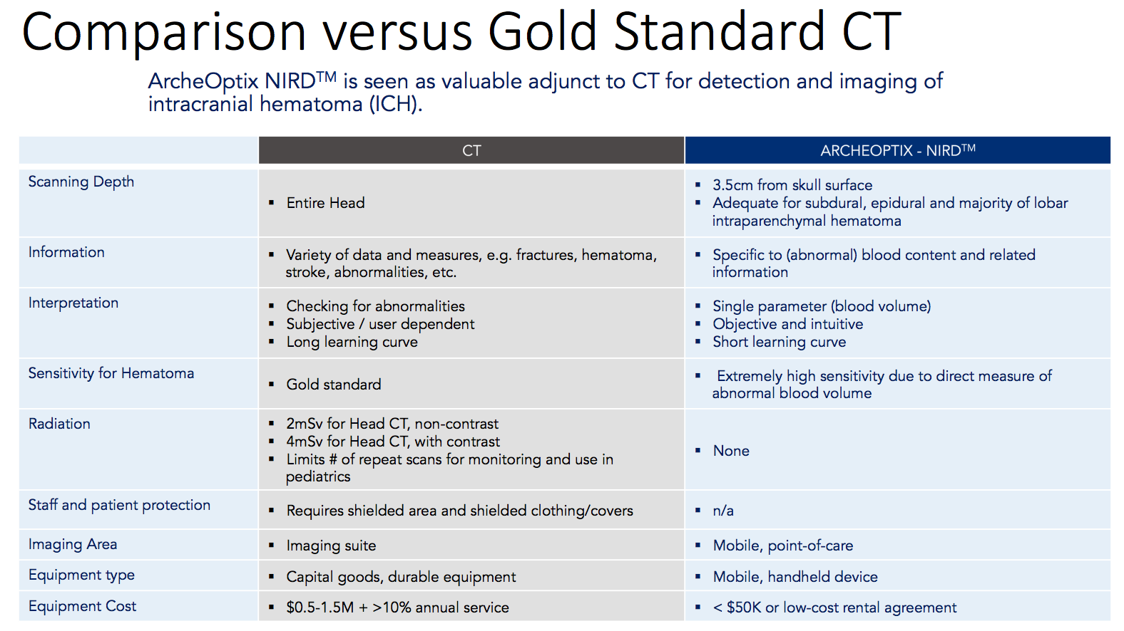
CT Scans vs. Our NIRD® System
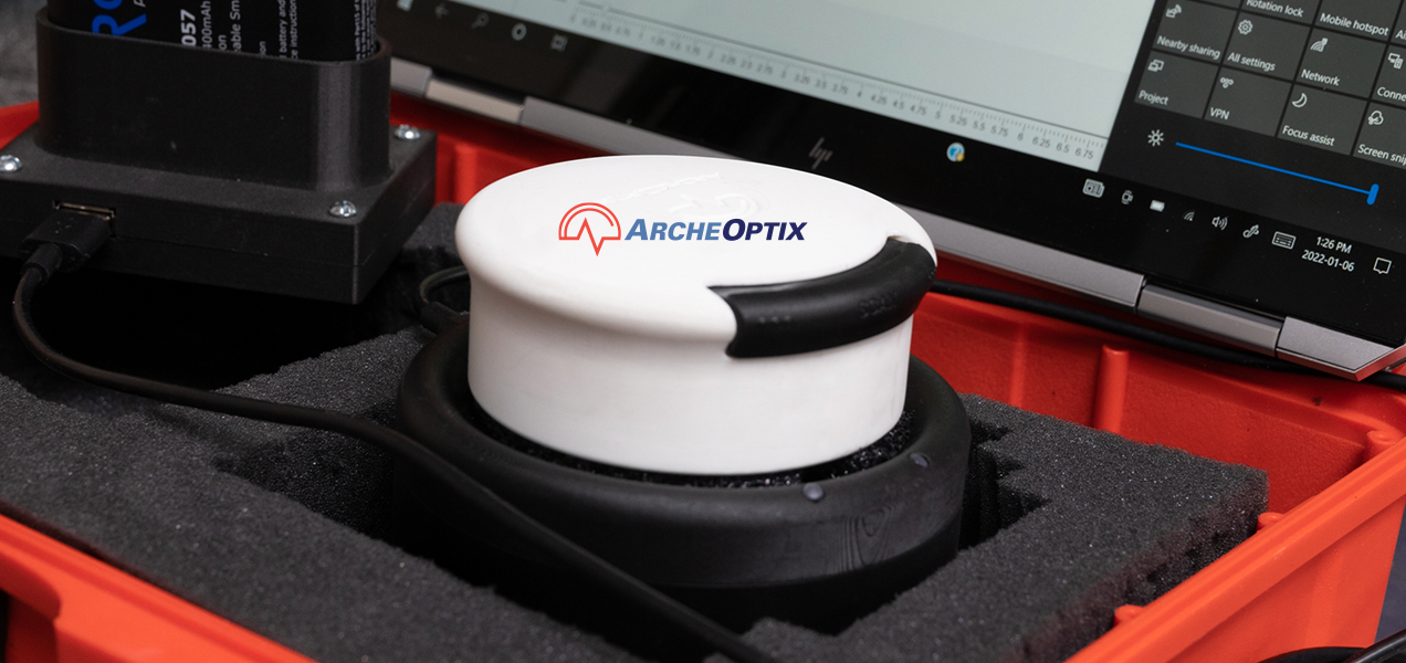
The NIRD® is a handheld portable imaging device, utilizing Near infrared technology with proprietary algorithms. Offers an instant brain image, anywhere.

Improved Patient Outcomes
Enhanced screening during TBI diagnosis at the point of Injury in remote settings
Monitoring at bedside
No radiation exposure
Provides a tool to frontline healthcare workers to more accurately assess the severity of head injury and determine required course of action
Reducing Inefficiencies in Health Care
- ~80% of CT scans determine that patient has no brain bleed
- Confirmation and prioritizing of first response
- Prioritizing the CT run-time and scheduling
- Reduction in unnecessary patient transport
- Potential for CT cost avoidance
- Free up healthcare resources that would typically be expended by transporting and assessing patients with CT scanners
Key Advantages
- No radiology expertise needed to identify bleeds at the site of injury
- CT Scan can be reserved for those who need it
Near Infrared Spectroscopy (NIRS)
The device scans at two separation distances (two depths). The near sensors track down to about 1cm depth, approximately to the inside of the skull – the far sensors track down to about 3.5 cm, into the intracranial space. The device is making a comparison measure between the near and far sensors as it tracks across the head (collecting 4000 data points per second). Effectively, the near sensors are expected to be tracking across “healthy” tissue, and the far sensors are looking for an abnormal pool of blood. Since blood absorbs light, there is a dramatic drop in the amount of light that would enter the far sensors, as compared to the near sensors. We now have a significant difference measure and can reliably say that there is blood in the intracranial space (Subdural/Epidural, et. al).
There is no issue with the skull in the Near Infrared (NIR) spectrum, which can be an issue in other modalities (ultrasound). However, skin tone (melanin) does – darker skin requires more light than pale skin to reach the required penetration depths. To deal with this, a very quick and simple calibration is performed at the front of the head (NOT in hair) to adjust/set the light level based on skin tone, for optimum performance. The scan of the head can now begin – it is important to maintain contact with the scalp.
The device is designed to have a combing effect which aids in getting through hair and maintaining contact with the scalp – long, straight hair is not a problem. A large amount of curly hair can be problematic; textured hair (braids, dreadlocks, etc.) are exclusions. Other exclusions are head tattoos (absorb all the light) and bruising greater than 0.5 cm (in both cases, you can try to navigate around them as you scan).
